L'Oréal Dermatological Beauty Pro is a community full of continuing medical education, patient resources, medical events, and more that support and empower healthcare professionals in their daily practice of dermatology.
Trichoscopy of dark-skinned SCALP
Pr. Bianca Maria Piraccini
Dermatologist, Researcher and Professor at the University of Bologna, Italy
- 6min (reading time)
- May. 2022
- Author: Pr. Bianca Maria Piraccini – Supported by

News on trichoscopy "4"
The dark-skinned scap is characterized by distinct features of the hair and the scalp and the absence of some specific signs of alopecia that are visible in white patients. This makes it difficult to evaluate pigmented patients on trichoscopy. Moreover, the incidence of some forms of alopecia is higher in dark-skinned compared with white patients (e.g. traction alopecia and hair damage due to cosmetic procedures, and central centrifugal is exclusive of pigmented scalp). In African Americans, the hair tends to be dryer, large in diameter, tightly curved and flat or elliptical in shape. The shaft tends to break easily, showing knots and longitudinal fissures.
(Trichoscopy of Dark Scalp. Ocampo-Garza J, Tosti A. Skin Appendage Disord. 2018 Nov;5(1):1-8. doi: 10.1159/000488885. https://www.ncbi.nlm.nih.gov/pmc/articles/PMC6323385/)
Normal scalp
The color of normal scalp in dark skinned patients may vary from light brown to black. It is quite common to see mild desquamation, asteriskc-like macules and residue of styling products, with one terminal hair o two vellus hair emerging from one hair follicle. The honeycomb pattern describes perifollicular hyperchromic lines corresponding to normal rete ridges melanocytes, interspersed with hypochromic areas corresponding to suprapappillary epidermis (Fig 1). Pinpoint white dots are small white dots corresponding to eccrine swear ducts openings. They show a regular distribution, like a “starry sky”, and they must be differentiated from cicatricial white dots (which is usually not regularly distributed, and shows larger white areas in addition to dots). The perifollicular halo: it is a white halo corresponding to the perifollicular epithelium.
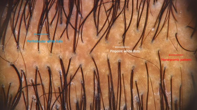
Figure 1. The normal black scalp.
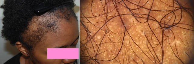
Traction alopecia (TA)
TA is very common in African Americans, affecting about one third of the population. It is typical of women, due to cultural hairdressing practices and to the curly shape, a reduced tensile strength and minor breaking point of the hair shafts. The typical wearing of high-tension hairstyles (e.g. buns, ponytailsfor, braids, dreadlocks, and hair extensions) for a prolonged time, leads to mechanical hair damage and inflammation of the hair follicle.
Hair loss is commonly observed frontal and temporoparietal areas, but it can affect the whole scalp, depending on the areas subject to traction. The damage may is reversible, but, if prolonged, it may lead to a permanent hair loss.
The clinical history of the patient is highly important in TA diagnosis, in addition to trichoscopy.
In the early stages, trichoscopy may reveal reduced hair density with several miniaturized hairs and also hair casts (Fig. 2), broken hair fragments, erythema and pustules. Late stages may present white patches and pinpoint white dots with an irregular cicatricial pattern.
(Traction alopecia: the root of the problem. Billero V, Miteva M. Clin Cosmet Investig Dermatol. 2018 Apr 6;11:149-159. https://www.ncbi.nlm.nih.gov/pmc/articles/PMC5896661/)
Figure 2. Traction alopecia.
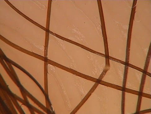
Alopecia due to hair breakage
High-tension hairstyles, in addition to the mechanical hair features of dark-skinned individuals, may lead to hair fragility and breakage. Patients may complain a lack of hair growth, but the tug test would confirm the presence of hair fragility. Hair brakage may determine also alopecic areas.
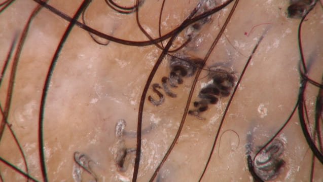
On trichoscopy, the usual signis are trichorrhexis nodosa, with white nodules among the shaft, lacking of cuticle and determined by a breakage of the hair shaft into multiple small fibrils, causing a segmental thickening in hair diameter. If hair shafts break, its ends present a peculiar brush-like appearance (Fig. 3).
(Hair Breakage in Patients of African Descent: Role of Dermoscopy. Quaresma MV, Martinez Velasco MA, Tosti A. Skin Appendage Disord. 2015;1(2):99-104. https://www.ncbi.nlm.nih.gov/pmc/articles/PMC4857843/)
Figyre 3. Trichorrhexis nodosa due to hair shaft traumatic damage.
Tinea capitis (TC)
TC is common in African American children. It is mainly caused by anthropophylic dermatophytes, inducing alopecia in multiple areas of the scalp, through endothrix invasion of the hair.
On clinical examination, the alopecic areas are covered by desquamation. Inflammation is present, too, but it might be hard to recognize due a scarce presence of erythema.
Typical trichoscopy findings that allow diagnosis are: 1) corckscrew hairs: short, broken and multiply twisted hair (Fig. 4); 2) morse code hair, 3) black dots, 4) hair casts, 5) comma hair: similar to those seen in white patients.
Despite the utility of trichoscopy, mycology is the gold standard for an aethiologic diagnosis and for monitoring the effects of the antifungal therapy.
Figure 4. Corckscrew hair in black haired tinea capitis.
Alopecia areata (AA)
The clinical and trichoscopic presentation of this autoimmune disorder is similar between white and dark-skinned people. However, some typical trichoscopic signs may be less evident and hard to recognize in dark people, due to a reduced color contrast: for example yellow dots may be scarcely evident as they appear white, in groups of two or three, surrounded by a whitish circular halo, corresponding to empty follicular units.
The most evident AA trichoscopic findings in these patients are black dots, exclamation mark hairs, broken and dystrophic hairs.
Androgenetic alopecia
Androgenetic alopecia in dark-skinned people, compared with Caucasians, is less common and also harder to recognize on clinical and trichoscopic examination. Usually, honeycomb patterns is more evident and there are pinpoint white dots mimicking empty follicular openings. Variability of the hair diameter is tyipically present in more than 20% of the hairs, together with short and thin regrowing hairs in the frontal area.
Other signs, such as the peripilar sign, may not be visible in these patients.
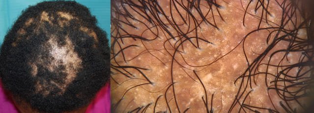
Central Centrifugal Cicatricial Alopecia (CCA)
CCA affects exclusively dark-skinned individuals, being the most common form of cicatricial alopecia in African young women. It is more rare in men, often misdiagnosed with androgenetic alopecia. It starts with an hair thinning of the vertex or central scalp (or with alopecic patches of the lateral and posterior scalp), progressing centrifugally during time. In early stages, it may resemble androgenetic alopecia, but it ends up in cicatricial alopecia.
Trichoscopy is not specific for this condition, showing reduced hair density, hair shaft variability, broken hair, black dots, absence of follicular openings, perifollicular scaling, and also single hair or groups of two hair surrounded by a peripilar gray-white halo (due to lamellar fibrosis surrounding the outer root sheath).
(Dermatoscopic features of central centrifugal cicatricial alopecia. Miteva M, Tosti A. J Am Acad Dermatol. 2014 Sep;71(3):443-9. https://www.jaad.org/article/S0190-9622(14)01427-3/fulltext)
Figure 5. Central Centrifugal Cicatricial Alopecia: single hair or groups of two hair surrounded by a peripilar gray-white halo.