L'Oréal Dermatological Beauty Pro is a digital community empowering healthcare professionals to improve their daily practice of dermatology through cutting-edge research, science and education on skin and hair care.

In vivo multiphoton FLIM quantification and characterization of melanins in European subjects with different variants in MC1R and SLC45A2 key pigmentation genes
E. Tancrède-Bohin, S. Brizion, S. Victorin, N. Parent, P. Bastien, N. Soufir, A.M. Pena
- 10min
- Jul. 2023
- Supported by

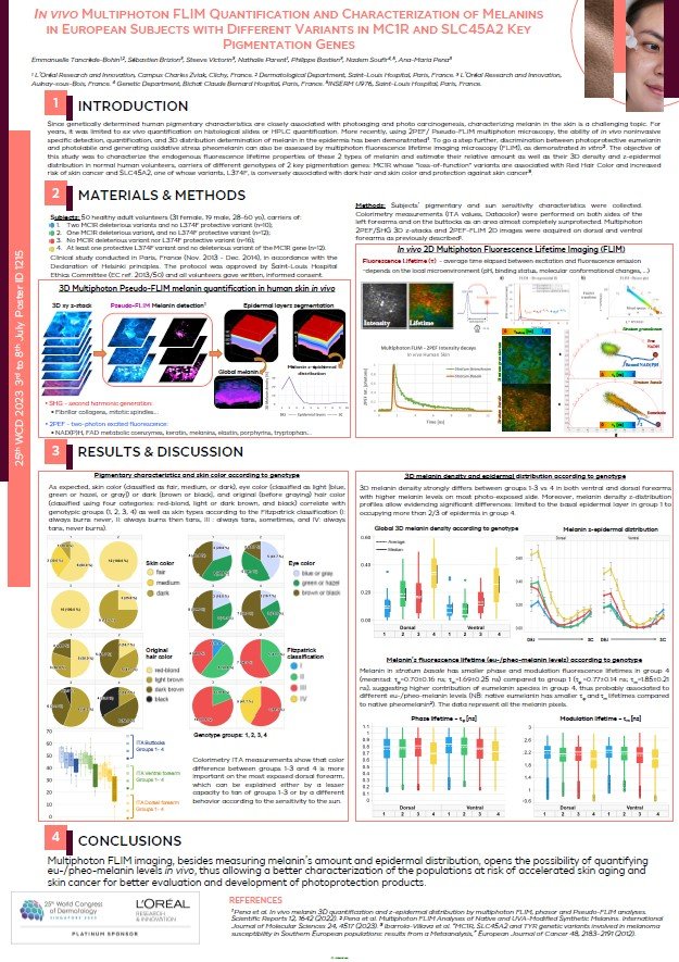
INTRODUCTION
Since genetically determined human pigmentary characteristics are closely associated with photoaging and photo carcinogenesis, characterizing melanin in the skin is a challenging topic. For years, it was limited to ex vivo quantification on histological slides or HPLC quantification. More recently, using 2PEF/ Pseudo-FLIM multiphoton microscopy, the ability of in vivo noninvasive specific detection, quantification, and 3D distribution determination of melanin in the epidermis has been demonstrated1. To go a step further, discrimination between photoprotective eumelanin and photolabile and generating oxidative stress pheomelanin can also be assessed by multiphoton fluorescence lifetime imaging microscopy (FLIM), as demonstrated in vitro. The objective of this study was to characterize the endogenous fluorescence lifetime properties of these 2 types of melanin and estimate their relative amount as well as their 3D density and z-epidermal distribution in normal human volunteers, carriers of different genotypes of 2 key pigmentation genes: MC1R whose "loss-of-function" variants are associated with Red Hair Color and increased risk of skin cancer and SLC45A2, one of whose variants, L374F, is conversely associated with dark hair and skin color and protection against skin cancer.